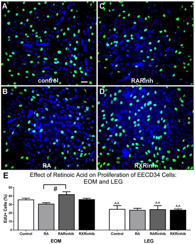
Fig. 5. EECD34 cell proliferation is unchanged by additional retinoic acid or reduced retinoic acid signaling.

Representative images of EECD34 cells from wild-type mouse EOM treated with (A) vehicle (control), (B) retinoic acid (RA), (C) the RAR inverse agonist BMS493 (RARinh), or (D) the RXR antagonist UVI 3003 (RXRinh) and stained for EdU (green) and DAPI (blue). (E) Percentage of EECD34 cells from EOM and LEG that have incorporated EdU following treatment with vehicle (control), retinoic acid (RA), BMS493 (RARinhib), or UVI 3003 (RXRinhib). Scale bar is 20 μm. # indicates significant difference from EOM EECD34 cells treated with retinoic acid ^^ indicates significant difference from EOM with the same treatment. Data are statistically significant at p ≤ 0.05.
