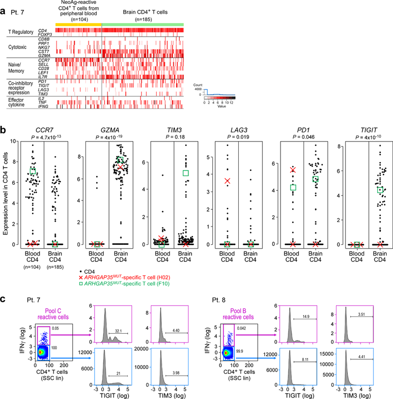
Extended Data Fig. 8 |. Comparison of single-cell expression profiles of circulating neoantigen-stimulated CD4+ T cells and tumour-associated CD4+ T cells isolated from patient 7.

a, Single-cell transcriptome analysis of CD4+ tumour-associated T cells (n = 185), freshly isolated at the time of relapse and neoantigen-reactive CD4+ cells isolated from post-vaccination (week 16) peripheral blood (n = 104) of patient 7 (excluding CD4 dropouts). Tumour-associated T cells expressed more granzyme A than circulating neoantigen-reactive CD4+ cells and showed higher expression of the co-inhibitory molecule TIGIT. b, Significantly altered expression of CCR7, GZMA, LAG3, PD1 and TIGIT was detected between CD4+ cells from the blood versus brain by two-sided Wilcoxon rank-sum tests. The expression levels of ARHGAP35MUT-specific single T cells (clones H02 (red cross) and F10 (green box)), as described in Fig. 4, are marked. c, Vaccination peptide pool-reactive T cells from patients 7 and 8 PBMCs were stained for TIGIT and TIM3, confirming the minimal expression of these markers on neoantigen-reactive IFNγ-producing T cells in the periphery, compared to IFNγ− controls, as suggested by the single-cell transcriptome data in a. Data are representative of results from two independent experiments.
