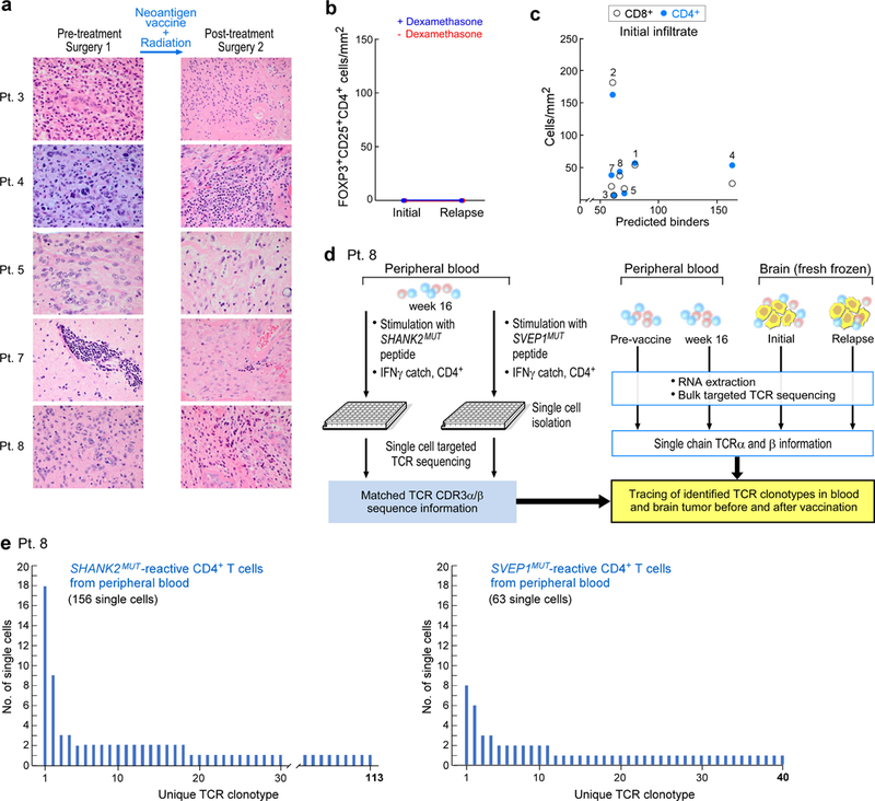
Extended Data Fig. 5 |. Analysis of tumour morphology, immune infiltration at initial resection and relapse, and TCR clonotypes of patient 8.

a, Haematoxylin and eosin staining of initial and post-vaccination resection samples show the most-prominent changes for patients 4, 7 and 8, including high levels of perivascular lymphocytes, extensive cystic changes and necrosis post-treatment in patient 7, low tumour content and patchy lymphocytic infiltration in patient 8 and sarcomatous morphology with myomatous changes in patient 4. b, No FOXP3+ and CD25+ CD4+ cells were detected in matched initial and relapse tumour sections, evaluated by multiplex immunofluorescence. c, No correlation was observed between the number of predicted neoantigens and the extent of T cell infiltration in the initial resection sample. d, Schema of single-cell TCR analysis of neoantigen-reactive T cells isolated from post-vaccination PBMCs of patient 8 and comparison to bulk TCR sequencing of cDNA from pre- and post-vaccination PBMCs and from initial and relapse fresh-frozen tumour biopsies. e, TCR clonotypes observed in neoantigen-reactive T cell lines generated from PBMCs of patient 8, based on single-cell-targeted TCRαβ sequencing (see Methods). This experiment was performed once, with the available resection tissue.
