
Fig 2. Heterogeneity and interactions among RCs.
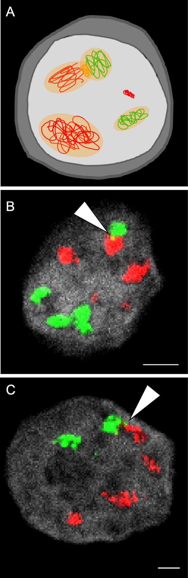
(A) Schematic illustration of an infected cell with two types of viral genomes (red or green lines) that form distinct RCs (RCs shown as orange area in the nucleus). Viral proteins within the RCs originate from the two genomes. Recombination events occur between two genomes in coalescing RCs (orange line). An entering genome that did not initiate an RC is also shown (condensed without background). (B, C) Fluorescence in situ hybridization image of U2OS (B) or Vero (C) cells 6 hours postinfection with two HSV-1 recombinants at MOI 20. Each viral recombinant carries a unique tag sequence that can be detected by a set of fluorescent probes (either green or red). Arrowhead points to a site of colocalization (indicating a possible recombination event). DNA staining was done by DAPI (gray). Scale bar, 20 μM. Experimental details were described [7]. MOI, multiplicity of infection; RC, replication compartment.