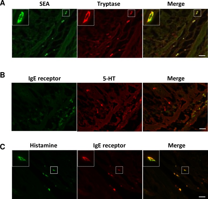
Fig 4. Characterization of SEA-binding mast cells.
Specimens of the intestinal tract were removed from a common marmoset. Frozen tissues were sectioned (10 μm) and incubated with SEA (1.0 μg/ml) for 60 min at room temperature. (A) Ileal sections were processed for double immunofluorescence staining using anti-SEA antibody (green) and anti-tryptase mAb (red), which recognizes the mast cell marker, tryptase. (B) Ileal sections were processed for double immunofluorescence staining using anti-FcεRIα antibody (green) and anti-5-HT antibody (red). (C) Ileal sections were processed for double immunofluorescence staining using anti-histamine antibody (green) and anti-FcεRIα antibody (red). Each scale bar is equal to 20 μm.

