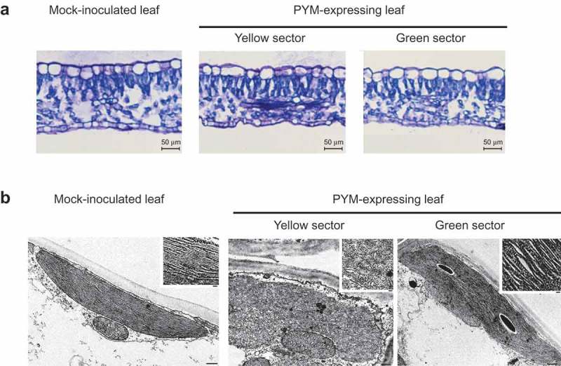
Figure 2.

Light and transmission electron microscopy (TEM) images of leaf sections and plastids of GF-305 peach seedlings. (a) Transversal sections of a leaf from a mock-inoculated plant and from the yellow and green sectors of a leaf infected by variant PYM-y4. Layers corresponding to epidermal, columnar palisade and spongy mesophyll cells are visible in the three instances. In the symptomatic leaves the sections from the yellow and green sectors appear slightly thinner than that from the mock-inoculated control. (b) TEM of mesophyll cells of a PYM-expressing leaf shows in the yellow sectors irregularly shaped plastids with thylakoids without the characteristic organization in grana and inter-grana, while such ultra-structural organization is preserved in plastids from the green sectors of the same leaf, in which the thylakoid interspaces are moderately larger than in the mock-inoculated control (bars 500 nm). Insets show magnifications of the respective thylakoids (bars 100 nm).
