. 2019 May 24;8:e45342. doi: 10.7554/eLife.45342

Figure 6. Delayed depletion of S100a4+cells impairs restoration of mechanical properties and alters matrix deposition.

(A) WT and S100a4-TK mice were treated twice daily with ganciclovir (GCV) from D5-10 post-surgery. (B) S100a4+ cell depletion results in a 91% reduction in S100a4 mRNA at D10 post-surgery (n = 3). (C) A substantial reduction in S100a4 protein expression was observed S100a4-TK repairs, relative to WT. Tendon is outlined in blue, scar tissue is outlined in black and (*) identify sutures (n = 4). (D–G) At D14 no change in MTP Flexion Angle (D) and Gliding Resistance (E) were observed between WT and S100a4-TK repairs. (F) Max load at failure was significantly reduced following S100a4-cell depletion, while no change in Stiffness was observed (G) (n = 7–10), (**) indicates p<0.01 (un-paired t-test). (H and I) Morphologically, (H) ABH/OG and (I) Picrosirius staining demonstrate reduced matrix deposition bridging the tendon ends in the S100a4-TK repairs, relative to WT. (*) Indicates sutures.

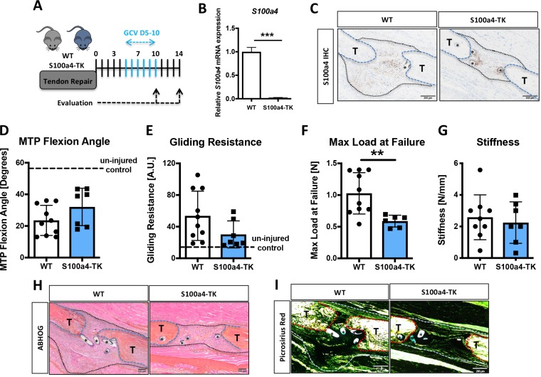
Figure 6.

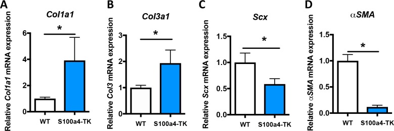
Figure 6—figure supplement 1. S100a4+cell depletion alters expression of matrix, tenogenic and myofibroblast-associated genes.

Figure 6—figure supplement 1.

qPCR analyses demonstrated significant increases in (A) Col1a1, and (B) Col3a1 expression, while (C) Scx and (D) α-SMA expression levels were significantly reduced in S100a4-TK, relative to WT. Data were normalized to expression in WT samples and the internal control β-actin. (*) Indicates p<0.05, (***) indicates p<0.001, (n = 3 per group) (un-paired t-test).
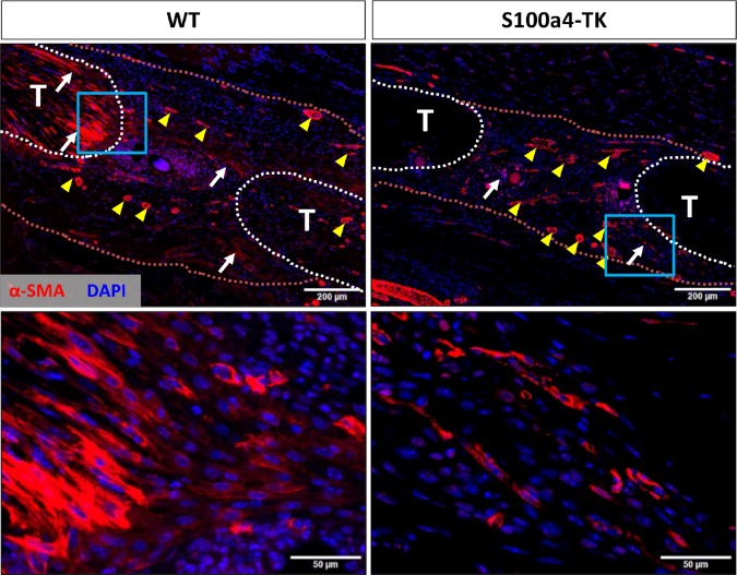
Figure 6—figure supplement 2. S100a4+cell depletion reduces α-SMA+myofibroblast content during healing.

Figure 6—figure supplement 2.

At D14 post-surgery, abundant α-SMA+ myofibroblasts (red) were observed in WT repairs. In contrast, α-SMA+ myofibroblast content was markedly reduced in S100a4-TK (D5-10) repairs. White arrows indicate areas of α-SMA+ cells in the healing tissue, while yellow arrowheads denote α-SMA staining of vessels. Tendons are outlined in white and scar tissue outlined in red.
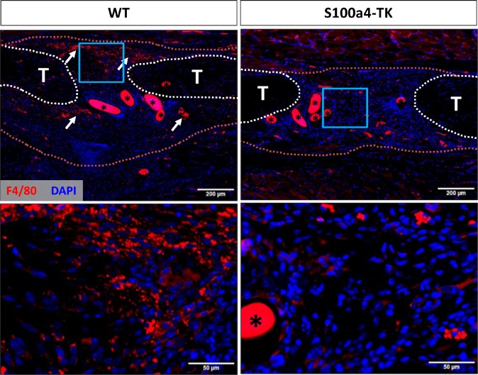
Figure 6—figure supplement 3. S100a4+cell depletion reduces macrophage content during healing.

Figure 6—figure supplement 3.

At D14 post-surgery abundant F4/80+ macrophages (red) were observed in WT repairs. In contrast, F4/80+ macrophage content was markedly reduced in S100a4-TK (D5-10) repairs. White arrows identify concentrated areas of macrophages. Tendon ends are outlined in white, while scar tissue is outlined in red. (*) indicates sutures.