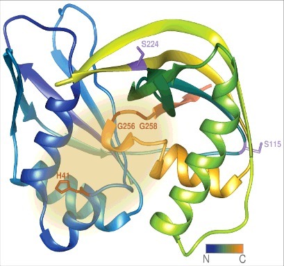
Figure 4.

The H. volcanii Cas6b protein. The structure of the H. volcanii Cas6b protein has not been solved experimentally, yet. Depicted is a structural model created by the Phyre 2 server [50], the suspected active site is highlighted in yellow. The amino acid residues coloured in red resulted in reduced crRNA levels when mutated to alanine. Position of His41 corresponds to the conserved active site histidine residues found across Cas6 species, whereas Gly256 and Gly258 are part of the glycine-rich loop implicated in crRNA positioning. The amino acid residues coloured in lilac correspond to those resulting in elevated crRNA amounts upon mutation to alanine (S115 and S224). They are located on the averted face of the protein in the analogous Cas6 of P. furiosus responsible for substrate binding. The N-terminus is coloured in blue and colour fades to orange reaching the C-terminus.
