
Figure 4. Vascular smooth muscle cell (VSMC)-specific SIRT1 (Sirtuin 1) overexpression prevents abdominal aortic aneurysm (AAA) formation and vascular pathophysiological mechanisms induced by angiotensin II (Ang II) infusion.
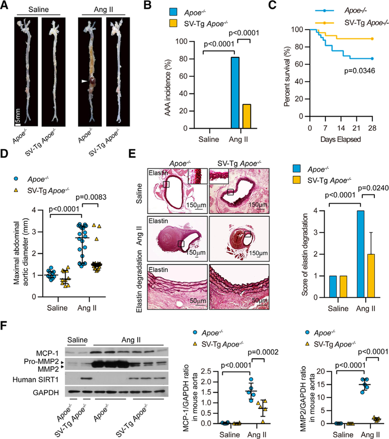
All mice were infused with saline or Ang II for 4 wk. A, Representative photographs showing macroscopic features of aneurysms induced by Ang II. The arrow shows a typical AAA (scale bars, 5 mm). B and C, The incidence (B) and survival curve (C) of Ang Il–induced AAA in SIRT1-VSMC–specific transgenic (SV-Tg) Apoe−/− mice (n=29) compared with those in Apoe−/− mice (n=33). There was no AAA formation in saline-infused mice (n=10), and the number of mice that developed AAA included the deaths caused by abdominal aortic rupture. D, The maximal abdominal aortic diameter in saline- and Ang II–infused mice. E, Representative staining with elastin and elastin degradation score in suprarenal aortas from saline- and Ang II–infused mice. The magnified photographs were taken at the location where the most severe elastin degradation occurred (scale bars, 150 and 50 μm; magnified photographs). F, Aorta homogenates were obtained from Apoe−/− and SV-Tg Apoe−/− mice infused with saline or Ang II for 4 wk. Western blot and densitometric analysis of the protein levels of monocyte chemoattractant protein-1 (MCP-1/CCL2) and matrix metalloproteinase 2 (MMP2) in aorta homogenates (n=4–6).