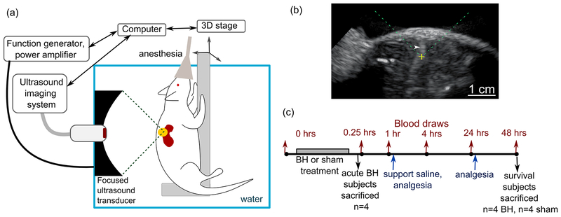
Figure 1.
(a) Schematic diagram of the experimental setup for ultrasound-guided boiling histotripsy (BH) ablation of renal tumors in Eker rats. (b) An example of B-mode ultrasound image during BH treatment. The position of the focused ultrasound (FUS) transducer focus is indicated on the screen as a yellow cross; the FUS beam is incident from the top of the image (dashed green lines). The transient elongated hyperechoic region (white arrowhead) that appears after each BH pulse corresponds to highly reflective vapor bubbles. (c) Timeline of the experimental procedures.

