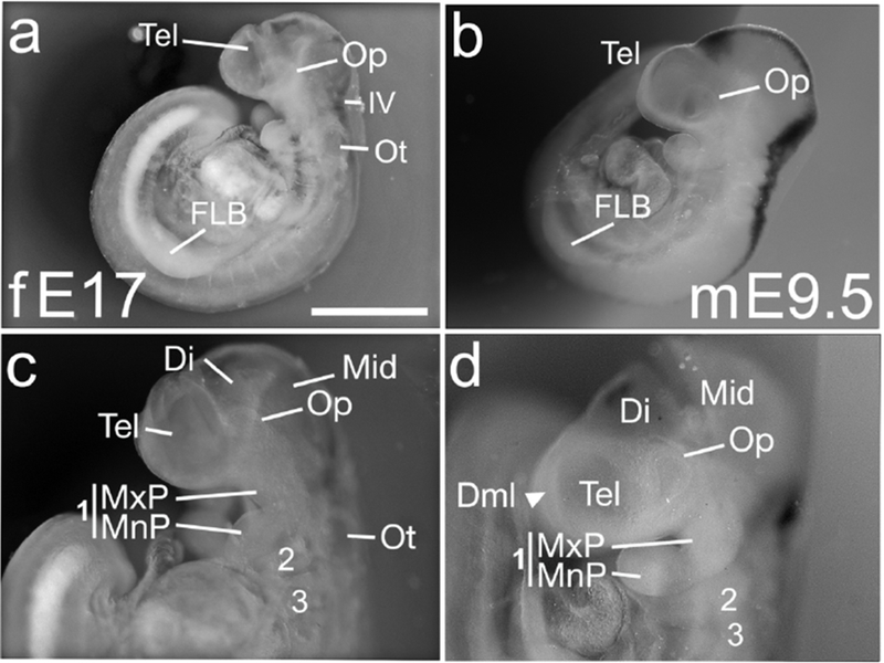
Figure 1. E17 ferret and E9.5 mouse, neural tube is closing and CNS structures are well defined.

(a-d) Lateral views of E17 ferret (fE17) (a,c) and two E9.5 mice (mE9.5) (b,d). (a,c) Ferret embryo, 29 somites. Rostral neuropore is closed (a,c). Three pharyngeal arches (1,2 and 3) are evident, as are the maxillary and mandibular prominences of the first arch (MxP, MnP) (c). The embryo has a forelimb bud condensation (FLB) (a), and the optic and otic vesicles (Op, Ot) are closed (a,c). The telencephalon (Tel), diencephalon (Di), midbrain (Mes) and the fourth ventricle (IV) are all distinct (a,c). (b,d) An E9.5 mouse, 29 somites, exhibits the same features. The telencephalon of a second E9.5 mouse exhibits a dorsal midline (Dml), which will ultimately separate the two cortical hemispheres (d, arrowhead); the ferret telencephalon (c) does not. Black staining in (b, d) is from ISH not relevant to the figure. Scale bar in (a) is 1.0 mm for a, b; 0.5mm for c, d.
