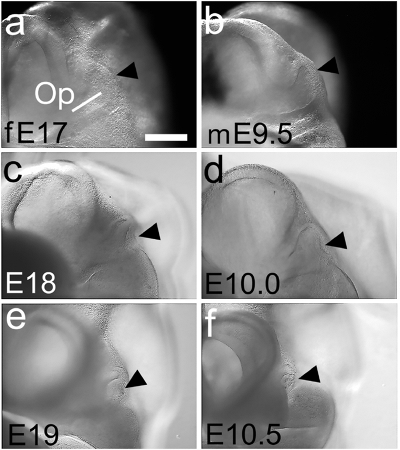
Figure 3. Optic vesicle development as a staging feature.

(a-f) Frontal views of three ferret and three mouse embryos at the ages marked. (a,c,e) The ferret Op epithelium has a smooth, slightly convex distal surface at E17 (a, black arrowhead), which progressively indents (c) and almost closes by E19 (e). (b,d,f) The mouse Op shows a similar progression (black arrowheads in b,d,f). Scale bar in (a) is 0.2mm for a,b; 0.3mm for c,d and 0.4 mm for e,f.
