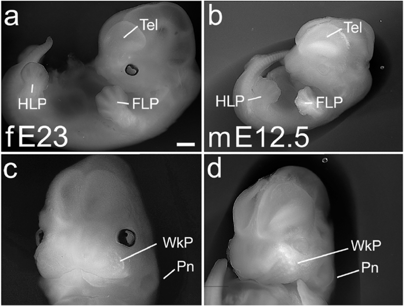
Figure 6. E23 ferret and E12.5 mouse, prominent development of face.

Whole embryo (a,b) and frontal head views (c,d) of an E23 ferret (a,c) and E12.5 mouse (b,d). The ferret at E23 shows accelerated overall growth compared with the mouse (a,b), but mouse and ferret share striking facial development. The facial prominences have coalesced, and both species show a prominent maxillary whisker pad (WkP) with rows of individual whisker primordia (c,d). The pinna of the ear (Pn) is also beginning to form (c,d). Eyes are pigmented in the ferret, but not in the albino mouse embryos (a,b). Scale bar in (a) is 1.0 mm in a,b, and 0.7mm in c,d.
