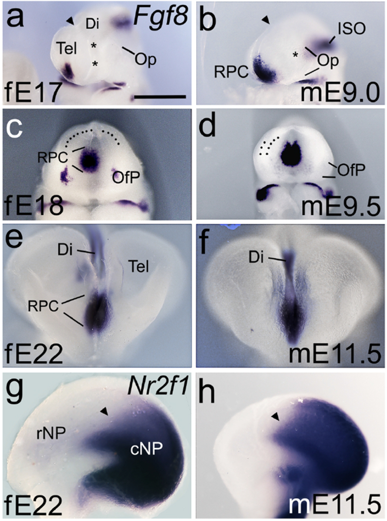
Figure 8. Fgf8 and Nr2f1 expression in the ferret and mouse rostral telencephalon.

(a-h) Whole brains or hemispheres viewed from the lateral side (a,b,g,h), rostral is left, or frontal views (c-f). (a-f) Fgf8 expression is less robust in the E17 ferret than in the E9.0 mouse (a,b), but strengthens by E18 to match the Fgf8 expression domain in the mouse at E9.5 (c,d). Asterisks in (a,b) indicate position of the optic vesicle (Op). Dotted lines in (c,d) indicate inner (c) or both inner and outer surfaces of the NP. Rostral Fgf8 expression domains remain similar in ferret and mouse at later ages (e,f), compatible with FGF8 involvement in later steps of forebrain development. (g,h) Expression of Nr2f1, repressed by FGF8, is confined to a similar caudal domain of the NP in E22 ferret and E11.5 mouse. In both, Nr2f1 expression has a relatively sharp rostral border (arrowheads in g,h). Scale bar in (a) is 0.5mm for a, b, 0.65mm for c – f, and 0.8mm for g,h. Abbreviations: ISO, isthmic organizer; rNP, rostral NP; cNP, caudal NP. Note difference in rostrocaudal length between fE22 and mE11.5 in (g,h).
