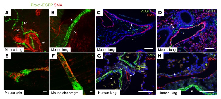
Figure 1. Pulmonary collecting lymphatics lack SMC or pericyte coverage.
(A and B) Whole-mount images of lungs from adult Prox1-EGFP lymphatic reporter mice show pulmonary lymphatic vessels (lv) in green, with the asterisk in B indicating an area of Prox1hi endothelial cells that marks lymphatic valves. Note the staining for SMA (red) present on both the bronchi (br) and arteries (art). (C and D) Immunohistochemical analysis of lung sections shows pulmonary lymphatics in green using staining for VEGFR3 or Lyve1 (arrows). Staining for SMA (C, red) or NG2 (D, red) marks airways and blood vessels, respectively. Asterisks indicate the large airway (C) and blood vessel (D) in proximity to lymphatic vessels. (E and F) Whole-mount staining for SMA (red) on lymphatic vessels in skin (E) and diaphragm (F) from Prox1-EGFP mice. (G and H) Human lung tissue sections were stained for the lymphatic molecular marker PDPN using the D240 antibody (red, arrows) and SMA (green), with arteries indicated by asterisks. Scale bars: 25 μm.

