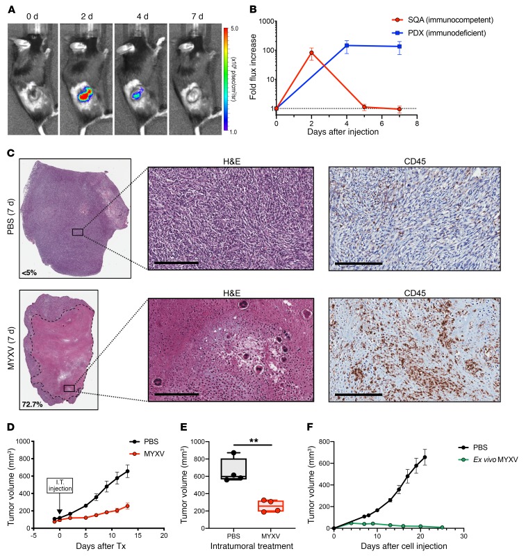
Figure 7. MYXV infection inhibits tumor development and Intratumoral delivery of MYXV to murine syngeneic SCLC allografts show necrosis, immune cell infiltration, and tumor growth inhibition.
(A) Subcutaneous allograft (SQA) SCLC tumor in an immunocompetent mouse showing replication and rapid clearance by 7 days after vMyx-FLuc treatment. (B) Quantification of the fold increase in vMyx-FLuc bioluminescence signal from SQA tumors in immunocompetent mice (n = 4) compared with PDX tumors in immunodeficient NSG mice (n = 3) following direct intratumoral injection of MYXV (vMyx-FLuc, 5 × 10⁷ FFU in 50 μl PBS). Data represent mean + SD. (C) Histological analysis of representative H&E-stained FFPE sections from SQA tumors 7 days after intratumoral injection of vMyx-FLuc (5 × 10⁷ FFU in 50 μl PBS) and 7 days after PBS injection (50 μl PBS). Extensive necrosis is observed in representative MYXV-treated tumor. Higher magnification illustrates the necrotic debris field with select immune cells further enlarged to observe nuclear morphology. Anti-CD45 IHC confirms high levels of immune cell infiltration following MYXV treatment. Scale bars, 200 μm. (D and E) Effect of either intratumoral injection of MYXV (vMyx-FLuc, 5 × 10⁷ FFU in 50 μl PBS) or PBS (50 μl). (D) Effect on tumor volume over time (n = 4 mice in each group). Data represent mean + SEM. (E) Final tumor volume at experimental endpoint (n = 4 mice in each group). Box-and-whisker plot showing the minimum to maximum with all data points and the horizontal line representing the median. **P < 0.01, by unpaired Student’s t test. (F) Murine SCLC cell line (737274-A) infected with vMyx-M135KO-GFP for 24 hours at 10 MOI (ex vivo infection) was injected into immunocompetent mice (n = 3) and monitored for tumor development then compared with control PBS-treated tumor development (n = 4). Data represent mean + SEM.

