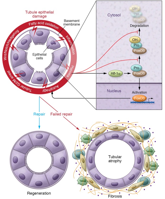
Figure 1. Failed tubule repair after injury leads to fibrosis.
Schematic diagram illustrating changes following tubule injury. A series of pathways leading to either regeneration or fibrosis includes failed completion of the cell cycle (G2/M arrest), activation of developmental pathways (Wnt/Notch/Hedgehog), and altered metabolism, including a defect in fatty acid oxidation, inflammation, and hypoxia. Hypoxia plays a pivotal role and a final common pathway in fibrosis. Hif activates FoxO3 expression through Hif1-α and inhibits FoxO3 prolyl hydroxylation and FoxO3 degradation. FoxO3 activation and accumulation can trigger tubular epithelial autophagy, which also plays a role in tubule injury. In contrast to the regeneration following injury, a failed repair of tubules after injury leads to tubule atrophy, an influx of inflammatory cells, and extracellular matrix accumulation, leading to the development and progression of fibrosis. Pro, proline residue.

