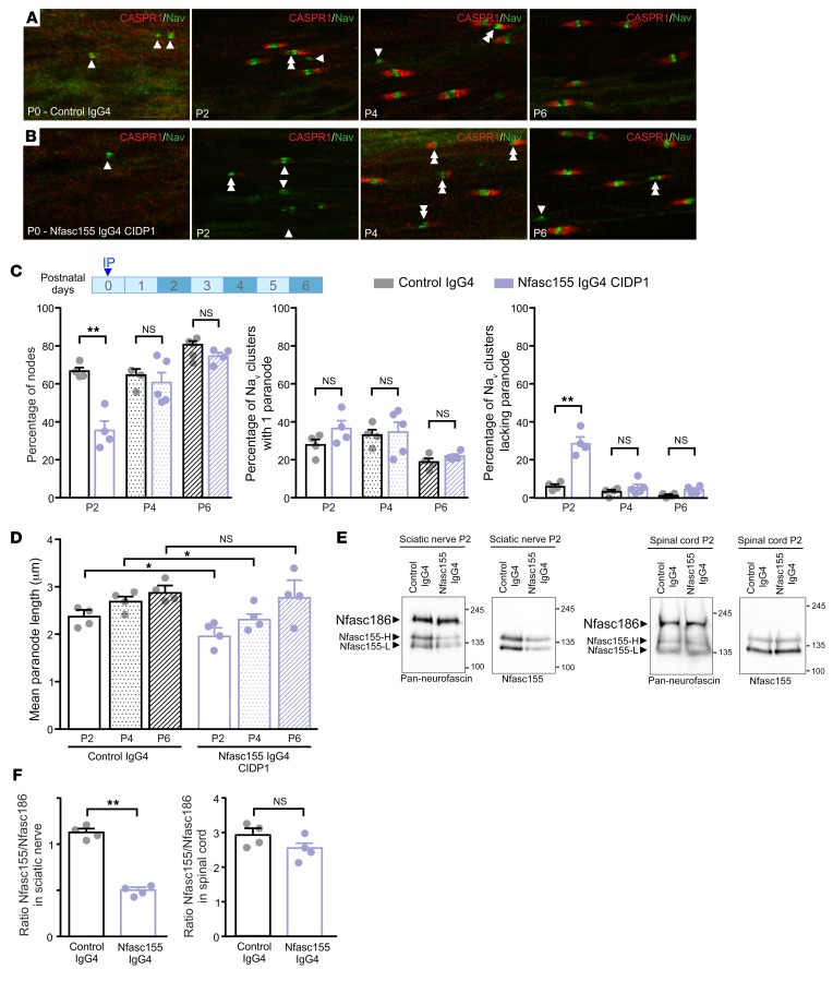
Figure 3. Passive transfer of anti-Nfasc155 IgG4 affects the formation of paranodal axoglial unit during development.
(A–D) Newborn rat pups received an i.p. injection of 250 μg of control IgG4 (A) or anti-Nfasc155 IgG4 from patient CIDP1 (B) on the day of birth (n = 4 animals for each condition and age). Sciatic nerve fibers were fixed and immunolabeled for voltage-gated sodium channels (Nav; green) and CASPR1 (red) at postnatal days 0 (P0), 2, 4, and 6. The percentages of Nav clusters with 1 or 2 flanking CASPR1-positive paranodes (double arrowheads) or without CASPR1-positive paranodes (arrowheads) were quantified at each age (C), as well as the paranodal length (D) (n = 200–300 nodes or paranodes for each condition and age). Injection of anti-Nfasc155 IgG4 importantly delayed the formation of CASPR1-positive paranodes, and a significantly higher percentage of heminodes without flanking paranodes was observed at P2 (**P < 0.005 by 1-way ANOVA followed by Bonferroni’s post hoc tests). Paranodal length was also significantly shorter 2 and 4 days after injection of anti-Nfasc155 IgG4 (*P < 0.05 by 1-way ANOVA followed by Bonferroni’s post hoc tests). Scale bar: 10 μm. (E and F) Sciatic nerve and spinal cord proteins (100 μg) from P2 animals injected with control IgG4 (n = 4) or anti-Nfasc155 IgG4 (n = 4) at P0 were immunoblotted with antibodies recognizing all neurofascin isoforms (Pan-neurofascin) or specifically Nfasc155. The level of Nfasc155 was quantified relatively to that of Nfasc186 in both sciatic nerve and spinal cord (F). Nfasc155 level was significantly decreased in sciatic nerves of animals treated with anti-Nfasc155 IgG4, but not in spinal cord (**P < 0.005 by unpaired 2-tailed Student’s t tests for 2 samples of equal variance). Molecular weight markers are shown on the right (in kilodaltons). Bars represent mean and SEM.

