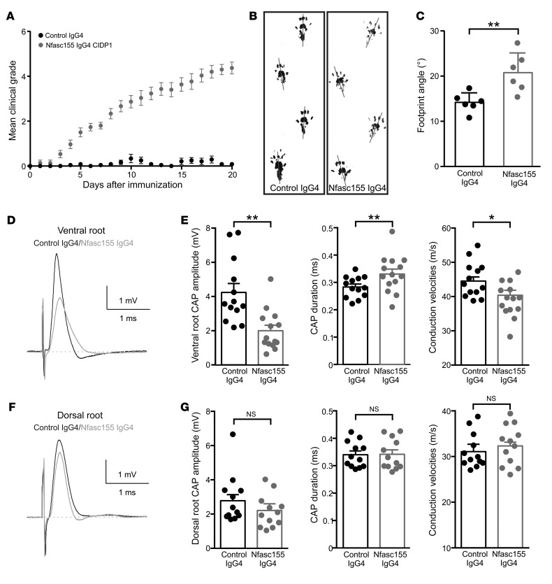
Figure 5. The chronic intrathecal infusion of autoantibodies induces gait abnormalities and motor nerve conduction slowing.
(A–C) Adult Lewis rats received daily intrathecal infusions (100 μg/d) of control IgG4 (black circles; n = 15 animals) or anti-Nfasc155 IgG4 from patient CIDP1 (gray circles; n = 15 animals) during 3 weeks. The clinical score was monitored daily and averaged. The passive infusion of anti-Nfasc155 IgG4 induced progressive clinical symptoms. (B and C) Footprint analysis revealed abnormal spreading of hind limbs in animals treated with anti-Nfasc155 IgG4 compared with control animals. The footprint angle (gray lines) was significantly increased in animals treated with anti-Nfasc155 IgG4 compared with controls. (D–G) L6 ventral and dorsal roots from animals injected with control IgG4 or anti-Nfasc155 IgG4 were recorded on day 21 after the beginning of the injections (n = 12–14 nerves from 12–14 animals). Representative CAPs from control IgG4–treated (black traces) and anti-Nfasc155 IgG4–treated (gray traces) animals are shown in D and F. The peak amplitude, CAP duration, and conduction velocities at peak amplitude are represented in E and G for ventral and dorsal roots, respectively. The ventral spinal nerves of animals treated with anti-Nfasc155 IgG4 showed a significant decrease in CAP amplitude and conduction velocity compared with controls. This was associated with a significant increase in CAP duration. By contrast, nerve activity was not significantly affected in dorsal root. *P < 0.05, **P < 0.005 by unpaired 2-tailed Student’s t tests for 2 samples of equal variance. Bars represent mean and SD.

