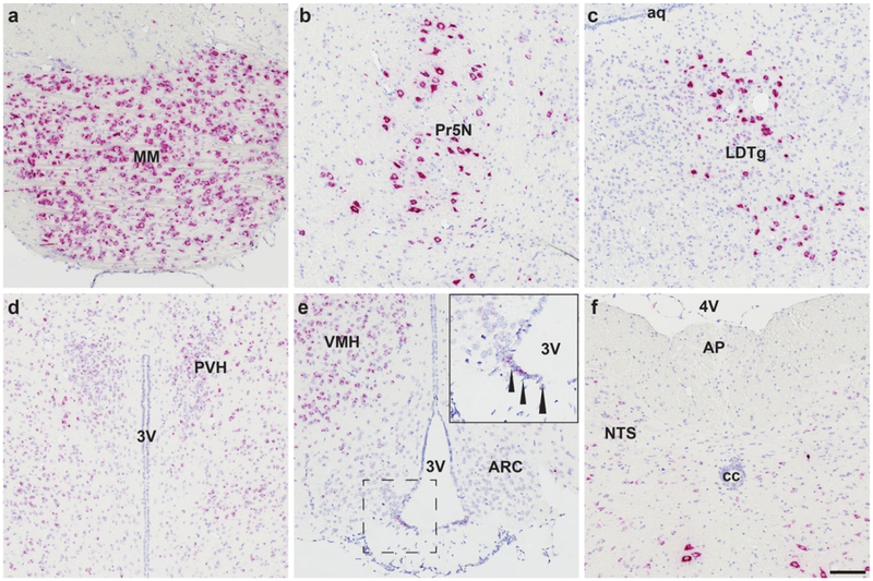
Figure 5.
Mm-Fgfr1c is expressed throughout the mouse CNS, with the most qualitatively robust expression observed in the mammillary body (MM, a), principal sensory nucleus of the trigeminal nerve (5N, b), and laterodorsal tegmentum (LDTg, c). These cells express so much Fgfr1 that the ISH staining appears to fill cell bodies, giving them a neuronal-like appearance. In food intake/energy balance governing centers, we observed consistent medium-high expression in the paraventricular hypothalamus (PVN, d), ventromedial hypothalamus (VMH, e), β2-tanycytes (inset, e), and nucleus of the solitary tract (NTS, f), but lower expression in the arcuate nucleus (ARC, e) or area postrema (AP, f). Scale bar = 200 μm.

