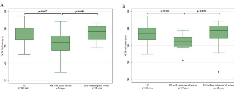
Figure 2.

Boxplots of GCIP thickness in HC and RIS by presence of spinal cord lesions (A) and infratentorial lesions (B)
GCIP: ganglion cell + inner plexiform layer; HC: healthy controls; RIS: radiologically isolated syndrome

Boxplots of GCIP thickness in HC and RIS by presence of spinal cord lesions (A) and infratentorial lesions (B)
GCIP: ganglion cell + inner plexiform layer; HC: healthy controls; RIS: radiologically isolated syndrome