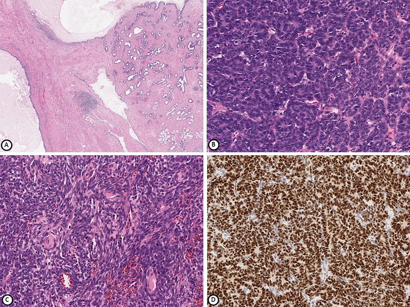
Figure 1.
Tumor from patient 1 comprised of mature teratomatous elements (A), blastemal and epithelial components (B) and spindle cell mesenchymal component (C). WT1 is positive in epithelial and blastemal cells (D). Hematoxylin-eosin stain (A-C), immunohistochemical stain (D); magnification x100 (A, B), x200 (C, D).

