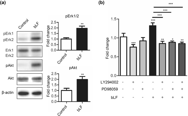
Fig. 2.
bLF increases phosphorylation of Erk and Akt in DP cells. a DP cells were serum-starved for 1 h and subsequently treated with 50 μg/mL of bLF for 1 h. Signals of phosphorylated proteins on Western blot analyses were quantified and normalized to their total protein levels. b DP cells treated with 50-μg/mL bLF and 5-μM LY294002, 5-μM PD98059, or a combined treatment of LY294002 and PD 98059 for 3 days. Cell proliferation was analyzed by the MTT assay. Data are presented as the mean values ± SD from three independent experiments. *p < 0.05; **p < 0.01; ***p < 0.001

