Fig. 3.
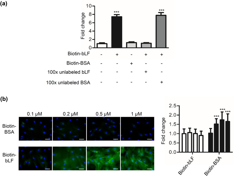
Binding of bLF to DP cells. a DP cells were treated with 0.1-μΜ biotin-labeled bLF or biotin-labeled BSA in the presence of 100-fold molar excess of unlabeled bLF or BSA for 4 h at 4 °C. The binding of biotin-labeled bLF to DP cells was detected by incubating them with HRP-conjugated avidin, followed by adding the OPD substrate reagent and measuring the absorbance at 492 nm. b Following methanol fixation and permeabilization, cells treated with biotin-labeled bLF or biotin-labeled BSA were stained for streptavidin-FITC Ab (green), and their nuclei were stained with DAPI (blue). Fluorescent microscopy showing DP cells treated with 0.1–1 μΜ of biotin-labeled BSA and biotin-labeled bLF for 1 h. The fluorescent levels of cells bound by biotin-bLF were quantified using AxioVision Software and normalized to the levels of biotin-BSA control. Scale bars, 50 μm. Data are presented as the mean values ± SD from three independent experiments. ***p < 0.001

