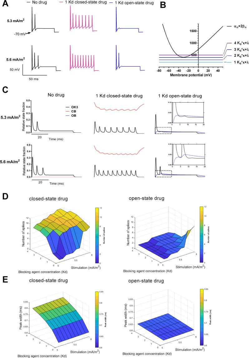
Figure 1.
Implications of differential state-dependent Kv channel block on the firing pattern. (A) Spiking patterns at 5.3 and 5.6 mA/m2 assuming no drug or 200 μM (corresponding to 1 Kd concentration) on a closed state binding model (red) and an open state binding model (blue). (B) Potassium channel closing (αn+2βn) and blocking rates (Lo · κ + λ) for 1–4 Kd equivalents as functions of membrane potential. (C) Potassium channel state fractions OK3 (black), CB (red) and OB (blue) at 5.3 and 5.6 mA/m2 assuming no drug or 200 μM (corresponding to a concentration of 1 Kd equivalent) on a closed state binding model and an open state binding model. Insets for the open state binding model demonstrate the accumulation of OB over time. (D) Number of action potentials (above −10 mV) during 60 ms at 5.1–6.0 mA/m2 stimulation for 0–800 μM (corresponding to 0–4 Kd equivalents) on a closed state binding model and an open state binding model. (E) Peak width of first action potential at −10 mV at 5.1–6.0 mA/m2 stimulation for 0–800 μM (corresponding to 0–4 Kd equivalents) on a closed state binding model and an open state binding model.

