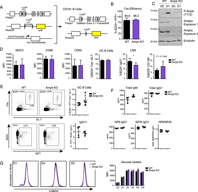
Figure 2.
Ampk deletion does not significantly impair B cell function. (A) Strategy for generating B-cell lineage specific knockout of Prkaa1 by crossing CD19-Cre recombinase (JAX: 006785) mice with Prkaa1fl/fl (JAX: 014141) mice and Rosa26 lox-STOP-lox YFP (JAX: 006148) mouse lines, yielding mice where CD19+ B cells lack the catalytic Ampkα subunit. (B) Quantification of Cre-recombinase efficiency by measurement of percentage of B220+ B cells that have YFP expression by flow cytometry in WT and Ampk KO B cells (n = 3). (C) Representative Western blot for P-Ampkα (T172), Ampkα and β-tubulin in WT and Ampk KO mice. Two exposures are shown for Ampkα to illustrate some remaining Ampk expression. Image was cropped for clarity, full-length blots/gels are presented in Supplementary Fig. 2. (D) Flow cytometry of WT and Ampk KO B cells during activation with anti-CD40L plus IL-4, including activation markers (MHCII, CD86, CD69) at 24 hours, GC differentiation (GC, %B220+ Fas+ GL7+) and CSR (%B220+ IgG1+) at day 3, and plasmablast differentiation (%B220lo CD138+) at day 5 (n = 5 for MHCII, CD86, CD69, and PB, n = 6 for GC, and n = 7 for CSR). (E) Flow cytometry of total splenocytes after immunization. Representative plots and quantification of GC differentiation (GC B Cells, B220+ Fas+ GL7+) and CSR (B220+ IgG1+) 14 days post-immunization with NP-(28)-CGG (n = 4). (F) Total IgM, total IgG1, NP8 IgG1, NP20 IgG1, and NP8/NP20 IgG1 ratio in serum 14 days after immunization of WT and Ampk KO mice with NP-(28)-CGG (n = 4). (G) Representative flow cytometry for 2NBDG glucose uptake at day 1, 3, and 5 post stimulation with anti-CD40 plus IL-4, and quantification at day 0 through 5 in WT and Ampk KO B cells (n = 3). Data represent mean ± SD (B,D–G). P values determined by Student’s t-test (B,D–G), *P ≤ 0.05.

