Fig. 6.
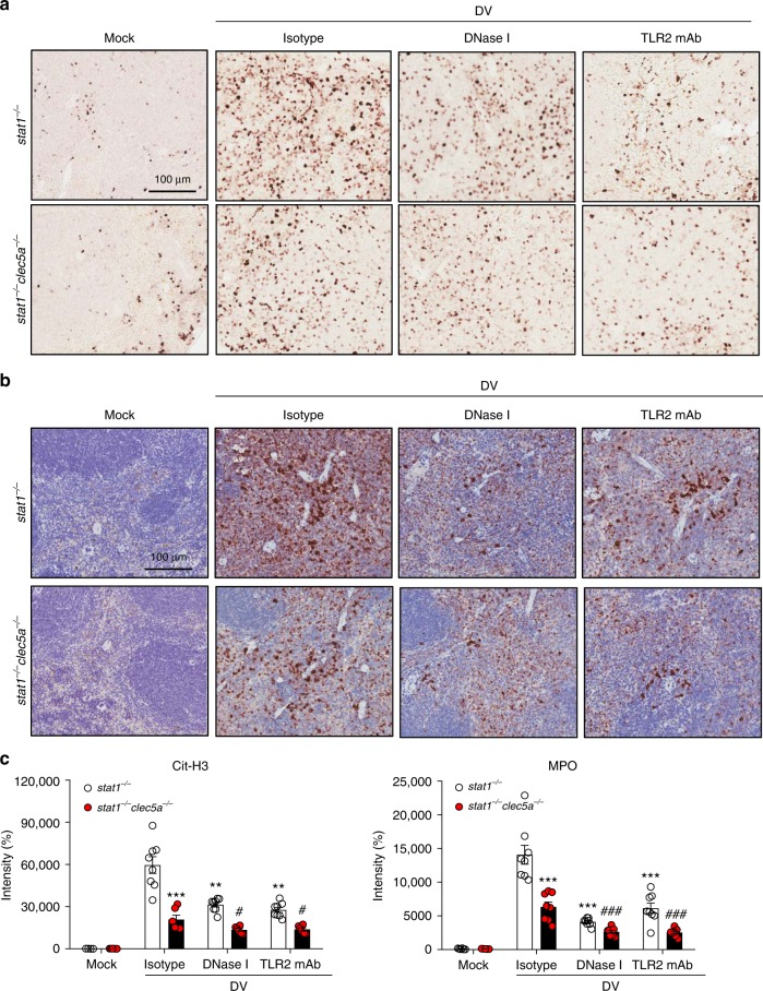
DV-induced NET formation in vivo occurs via CLEC5A and TLR2. a and b Stat1−/− and stat1−/−clec5a−/− mice were pre-treated with isotype control mAb, DNase I, and anti-TLR2 mAb, followed by inoculation with DV (NGC-N) via intraperitoneal route. Spleens were harvested and fixed at day 5 post-infection for immunohistochemical staining using anti-citrullinated histone H3 (Cit-H3) mAb (a) or anti-MPO mAb (b) (n = 3 per group). c Cit-H3 and MPO were quantified using MetaMorph image analysis software. **p < 0.01, ***p < 0.001 (compared with isotype control-injected stat1−/− mice); #p < 0.05, ###p < 0.001 (compared with isotype control-injected stat1−/−clec5a−/− mice) (Student’s t-test). Scale bar: 100 μm. Source data are provided as Source Data file

