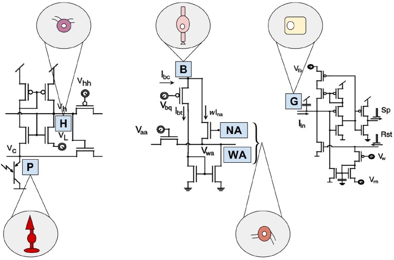
Figure 4.
Artificial building blocks and their biological models of the Parvo-Magno Retina from Zaghloul and Boahen. The left circuit shows the outer retinal layer. A phototransistor takes current via an nMOS transistor. Its source is connected to Vc, representing the biological photoreceptor (P). Its gate is connected to Vh, portraying horizontal cells (H). The circuit in the center represents the amacrine cell modulation. A bipolar terminal B excites a network of wide-field amacrine cells (WA) and also narrow-field amacrine cells (NA) through a current mirror. The amacrine cells, in turn have an inhibitory effect on B. By the right circuit the spiking ganglion cells are represented. Current Iin from the inner retinal circuit charges up a membrane capacitor G, based on biological ganglion cells. If its membrane voltage crosses a threshold a spike (Sp) is emitted and a reset (Rst) discharges the membrane. Inspired by Zaghloul and Boahen (2006).

