Figure 1.

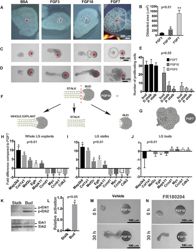
Effects of FGFs on LG and lung explants. (A) Different FGFs induce different lung airway dilation. Heparin sulfate beads were loaded with different FGFs or BSA (control) and implanted into the central part of each lung explant (isolated from mouse embryos at E12.5). FGF3 forms a much sharper gradient than FGF10 and induces lung dilation at a shorter distance from the bead, while FGF7 diffuses more freely inducing dilation within a large lung area. (B) Graphical representation of lung dilation shown in (A) p < 0.01, n = 10. (C,D) Different FGFs induce different morphology of lung (C) and LG (D) epithelial explants. Lung and LG epithelial explants exposed to FGF10 migrate towards the bead and have defined “bud” and “stalk” morphology, while FGF3 have a well-formed “stalk” but almost no “bud.” Both LG and lung explants exposed to FGF7 show extensive growth and formation of enlarged buds but not stalks (Beads are shown with red asterisk). (E) Quantification of BrdU labeling in different regions of the LG explants exposed to FGF3, 7, and 10 ligands. Explants exposed to FGF3 showed no significant increase in cell proliferation within the bud area, whereas explants exposed to FGF7 showed increase in cell proliferation throughout the whole explant. Application of the FGF10 induced cell proliferation only within the “bud” region. Quantification of proliferating cells was performed in four independent experiments (in 8–12 explants of each kind). “∗” marks significant difference between “D. stalk,” “P. stalk,” and “bud” regions. (F,G) Schematic diagram of experimental design. LG explants were exposed to FGF10 (F) or FGF7 (G) for 30 h and processed for qRT PCR. “Buds” and “stalks” areas of some explants exposed to FGF10 were separated mechanically and processed for qRT PCR. (H–J) Gene expression levels were examined by real-time RT-PCR custom array focused on the proliferation and differentiation markers in whole LG explants (H), stalks (I), and buds (J) of explants exposed to FGF10 and the expression profiles of these groups of genes were compared to that of FGF7 [shown as a 0 (zero) line]. “∗” marks significant difference in each gene expression compared to FGF7. (K) Extracellular signal-regulated kinases (ERK1/2) phosphorylation by FGF10 is significantly downregulated in “stalk” compared to “bud” areas of the LG epithelial explant. (L) Graphic representation of results (n = 3) shown in (K). “∗” marks significant difference in ERK1/2 phosphorylation between “stalk” and “bud” regions. (M,N) Selective inhibition of extracellular signal-regulated kinase ERK1/2 leads to lack of epithelial bud and decreased migration towards the FGF10 loaded bead.
