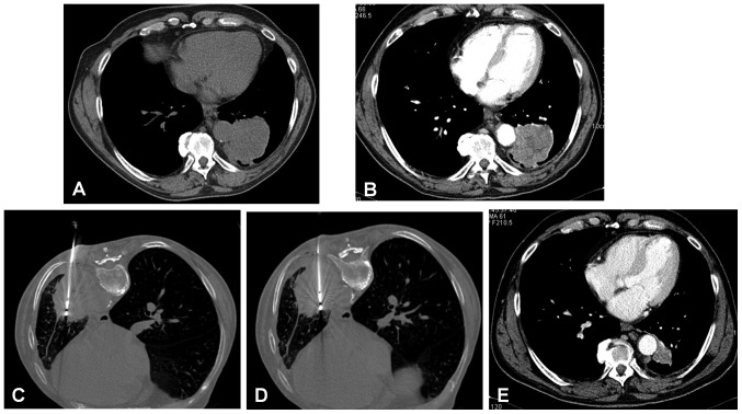
Figure 3.
MWA of squamous cell carcinoma (4×5 cm) of the left lung located in close proximity to the descending aorta in a 67-year-old man. (A) Basal unenhanced CT scan and (B) contrast-enhanced CT scan. (C) MWA by insertion of an antenna with output power of 70 W for 10 min with patient in prone position. (D) MWA by insertion of a second antennae with an output power of 70 W for 10 min with the patient in a prone position. (E) Follow-up CT scan at 10 months showing a tumor size reduction (1.8 cm) with subtle enhancement. MWA, microwave ablation.

