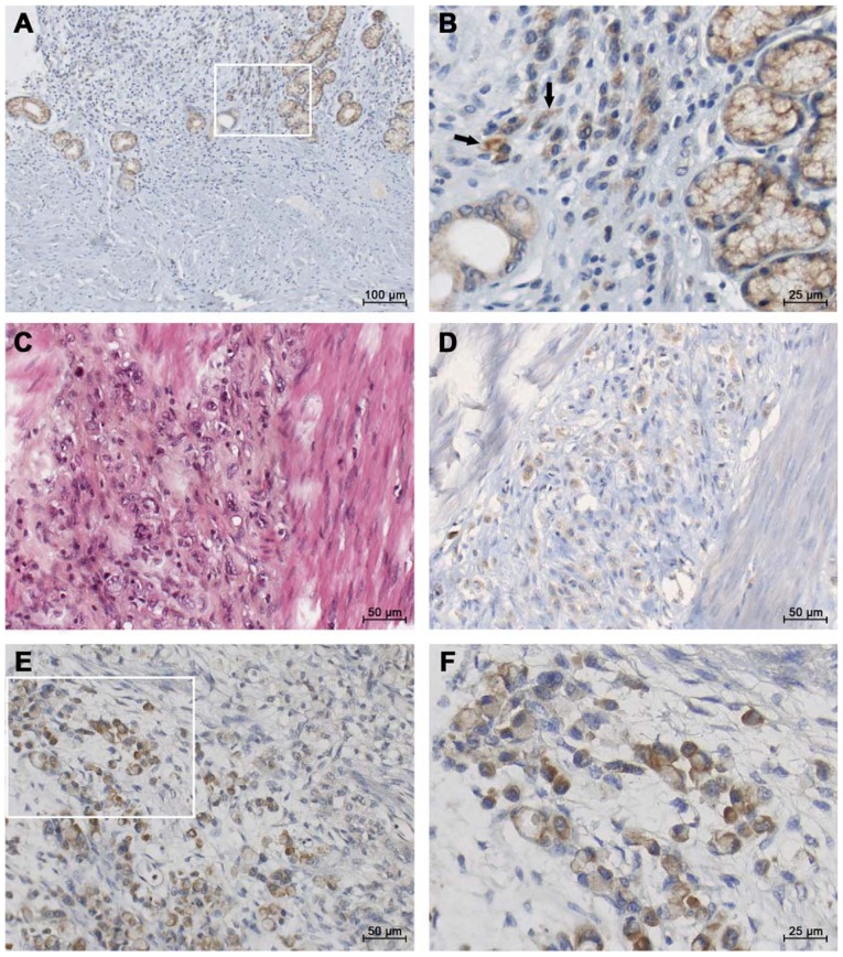
Figure 1.
Representative micrographs of IGF1 immunohistotochemical staining in diffuse-GC. Paraffin sections from gastric tissues were incubated with polyclonal antibodies against IGF1. The figure presents IGF1 immunostaining in diffuse-GC with different stages of tumor invasion. (A and B) IGF1 staining in glandular and independent epithelial cells of the gastric sub-mucosa of a diffuse-type GC (TNM2b); inset: higher magnification. Arrows indicate independent IGF1 positive tumor cells. (C and D) Aggressive diffuse-GC associated with fibrosis (linitis, TNM4) in a young patient; (C) hematoxylin-eosin staining and (D) IGF1 staining in invasive tumor cells. (E and F) Strong IGF1 immunostaining in a metastatic diffuse carcinoma; inset: higher magnification. GC, gastric cancer; IGF1, insulin-like growth factor 1.

