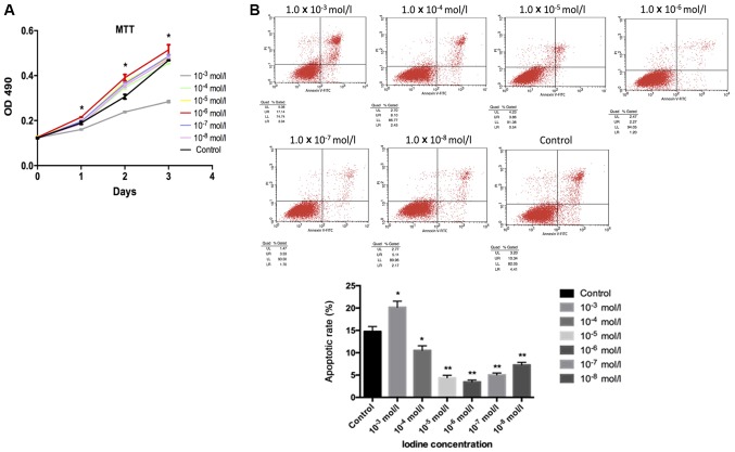
Figure 1.
Cell proliferation evaluated by MTT assay and flow cytometry. (A) It was observed that iodine at lower concentrations (1.0×10⁻⁴, 1.0×10⁻⁵, 1.0×10⁻⁶, 1.0×10⁻⁷ and 1.0×10⁻⁸ mol/l) contributed to the proliferation of BCPAP cells, while iodine of a higher concentration (1.0×10⁻³ mol/l) had a negative effect. Cells treated with iodine at 1.0×10⁻⁶ mol/l exhibited the most pronounced changes. (B) BCPAP cell apoptosis was analysed by flow cytometry with different concentrations of iodine. BCPAP cell apoptosis, when incubated with lower concentrations of iodine (1.0×10⁻⁴, 1.0×10⁻⁵, 1.0×10⁻⁶, 1.0×10⁻⁷ and 1.0×10⁻⁸ mol/l) was significantly inhibited. Among these, the highest inhibitory effect was observed with 1.0×10⁻⁶ mol/l iodine. A higher concentration of iodine (1.0×10⁻³ mol/l) appeared to promote cell apoptosis. *P<0.05 and **P<0.01 vs. the control group. OD, optical density.

