Figure 1.
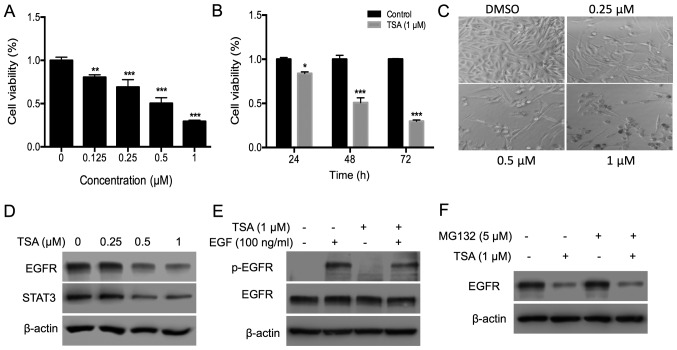
TSA inhibits PC3 cell proliferation by disrupting the EGFR-STAT3 pathway. (A) PC3 cells (3×10³) were plated in a 96-well plate and treated with different doses of TSA (0, 0.25, 0.5 and 1 µM) for 72 h; an MTT assay was used to measure the effects of TSA on PC3 cell proliferation. (B) PC3 cells were subjected to the aforementioned treatments, and the time-course effect of TSA (1.0 µM) was measured using an MTT assay. (C) Effects of TSA on the density and morphology of PC3 cells were observed via microscopy. The magnification is ×100. (D) Expression of EGFR and STAT3 in PC3 cells treated with TSA. PC3 cells (4×10⁵) were plated in 6-cm dishes, and 0.25, 0.5 or 1 µM TSA was added for 24 h. DMSO was added into the control group wells. Cells were collected and lysed with lysis buffer and the protein expression analyzed via western blotting. (E) TSA inhibits the phosphorylation of EGFR induced by EGF. Cells were starved in serum-free medium for 12 h and then treated with 1 µM TSA for 30 min, followed by EGF (100 ng/ml) for 10 min. Cells were collected, and western blotting was used to evaluate the phosphorylation of EGFR. (F) Expression of EGFR in PC3 cells treated with TSA and MG132. Cells were treated with 1 µM TSA and 5 µM MG132 for 24 h. Cells were collected, and western blotting was used to evaluate the expression of EGFR. *P<0.05; **P<0.01; ***P<0.001 vs. respective control. TSA, trichostatin A; EGFR, epidermal growth factor; p, phosphorylated; DMSO, dimethyl sulfoxide.

