Figure 3.
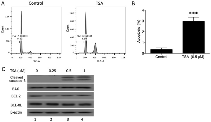
TSA induces apoptosis in PC3 cells. (A) Representative histogram of apoptosis induced by TSA in PC3 cells. (B) Proportion of apoptotic PC3 cells following treatment with TSA. ***P<0.001 vs. control. (C) PC3 cells were treated with different doses of TSA (0, 0.25, 0.5 and 1 µM), and then collected 24 h after treatment. Cleaved caspase-3, BAX, BCL-2 and BCL-XL were detected using Western blotting. TSA, trichostatin A.

