
Figure 1.
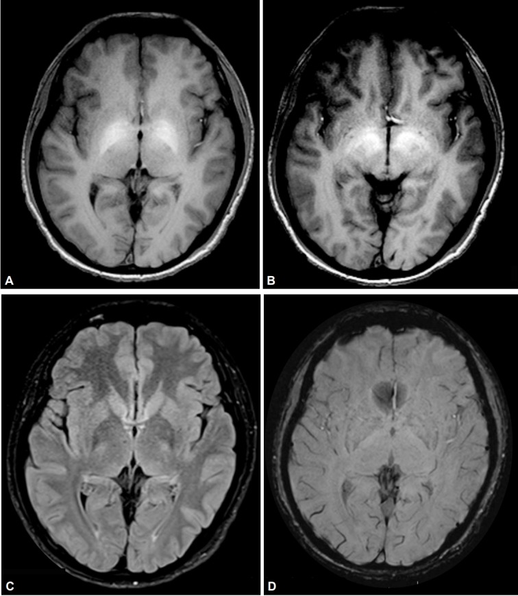
Magnetic resonance images demonstrating features suggestive of manganese toxicity. A and B: Axial T1-weighted images showing bilateral and symmetrical hyperintensities of the globus pallidus and substantia nigra. C: Normal T2 fluid-attenuated inversion recovery imaging. D: Normal susceptibility weighted imaging.