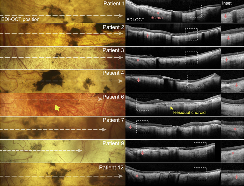
FIGURE 3.

Spectral-domain optical coherence tomography (SD-OCT, center column) with corresponding fundus regions (left column) of patients in the scleral exposure stage of Stargardt disease. Horizontal SD-OCT scans through the fovea reveal a complete loss of the reflective outer retinal bands, external limiting membrane, ellipsoid zone, interdigitation zone, and retinal pigment epithelium, as well as choroidal layers. Hypertransmission of the OCT signal is visible throughout the scan, revealing the underlying sclera (red arrowhead) in maginfied SD-OCT insets (right column). A central region of residual choroid (yellow arrow) was observed in Patient 6.
