Figure 5.
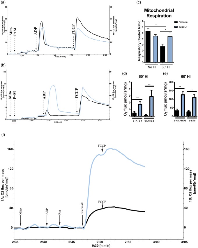
MgSO4 preconditioning preserved brain mitochondrial respiration after HI.(a) Respirogram showing vehicle (black) and MgSO4-(blue) treated samples analyzed 24 h post injection. (b) Respirogram showing vehicle (black) and MgSO4-(blue) treated samples analyzed after 30 min of HI. Respiration was significantly higher in the MgSO4-treated group compared to vehicle (p < 0.05) (Unpaired t-test, Mean ± SEM) (n = 6–7/group). (c) Mitochondrial respiration. A significantly higher RCR was found in brain mitochondria isolated from animals pretreated with MgSO4 compared to vehicle-treated animals after 30 min of HI (30′HI). No difference could be detected in RCR between MgSO4-treated animals with or without HI, whereas there was a ∼50% drop in RCR in response to HI in the vehicle-treated group (p < 0.002), (ANOVA, p < 0.05). (d) Basal mitochondrial respiration (STATE 1) as well as respiration in the presence of added ADP but absence of reducing substrates (STATE 2) in vehicle (black) vs. MgSO4 (blue; n = 4/group) (p < 0.03). (e) Complex-II-mediated respiration driven by succinate as reducing substrate during blockage of Complex-I by Rotenone in MgSO4 vs. vehicle (p < 0.03). Also, maximal ETS capacity induced by addition of FCCP (p < 0.03) (n = 5/group), (Mann–Whitney U test, Mean ± SEM). (f) Respirogram showing the respiratory activity of isolated mitochondria from MgSO4 (blue) compared to vehicle (black) group. The time-point for addition of each substrate is indicated by arrows in the figure.

