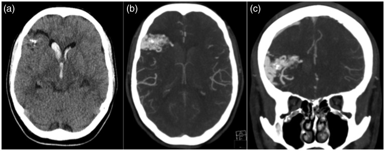
Figure 1.
(a) The unenhanced axial computed tomography (CT) scan demonstrates intraventricular haemorrhage with several foci of calcification within the right frontal lobe. A CT angiogram demonstrates a right frontal arteriovenous malformation with an aneurysm directed into the right frontal horn of the lateral ventricle that was believed to be the source of the haemorrhage (b and c).

