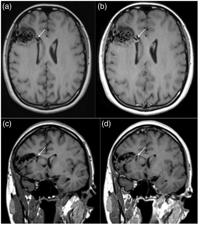
Figure 2.
A magnetic resonance image with pre and post-contrast T1-weighted sequences was performed. The aneurysm demonstrated on the computed tomography angiogram was clearly visible on the pre-contrast T1-weighted sequences (a and c, white arrow) with thick, circumferential enhancement of the aneurysmal wall seen on the post-contrast sequences (b and d, white arrow). There was no significant enhancement seen elsewhere within the arteriovenous malformation.

