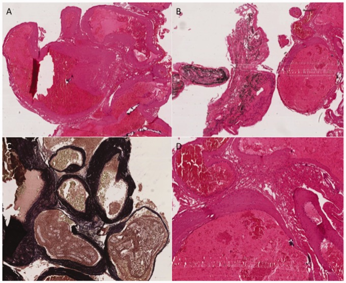
Figure 5.
Arteriovenous malformation demonstrating classic histological appearances of (a) closely associated, sometimes tortuous, ectatic vascular channels with walls of varying thickness and size. Occasional vessels contain black-staining embolisation material (Onyx) (b). Several vessel walls contain prominent internal elastic laminae, which are demonstrated on Masson trichrome staining as dark staining within the vessel walls (c). A higher power representative view demonstrating no histological evidence of mural inflammatory cell infiltrate within the vessel walls (d).

