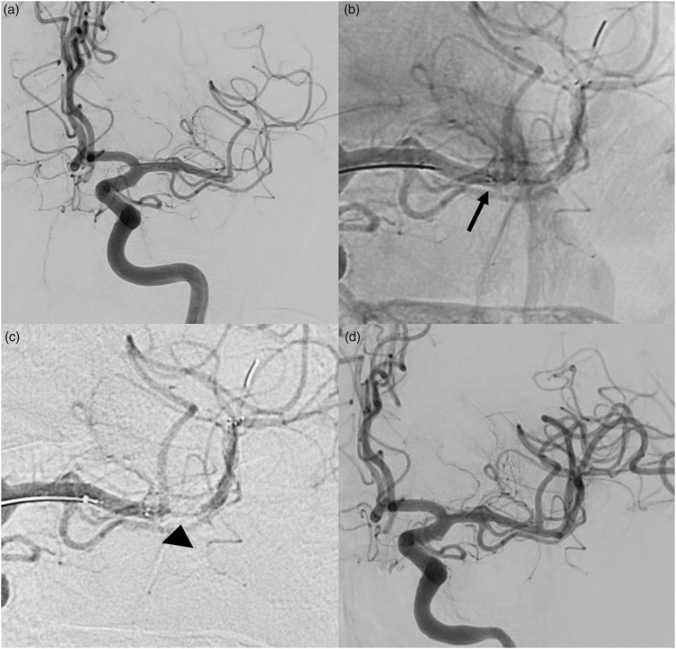
Figure 2.
(a) Left internal carotid artery (ICA) injection shows the embolic occlusion of the inferior branch (M2) of the left middle cerebral artery (MCA); (b) EmboTrap II opened with its proximal marker at the beginning of the clot (arrow); (c) left ICA injection shows a flow-channel within the EmboTrap II allowing immediate reperfusion of the ischaemic territory (arrow-head); (d) final angiogram showing the complete recanalization of the MCA.

