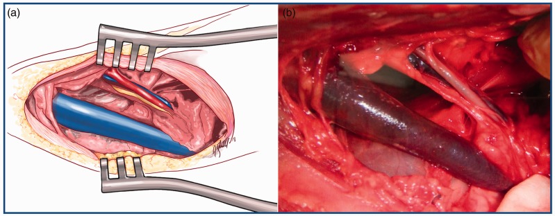
Figure 1.
displays a schematic drawing (image a) and a surgical picture (image b) of a cervical dissection and exposure of the neurovascular bundle of the neck. This is the site of implantation of the models. It includes the common carotid artery, the vagosympathetic trunk and the internal jugular vein. Of greater thickness, the external jugular vein is also exposed in the dissection.

