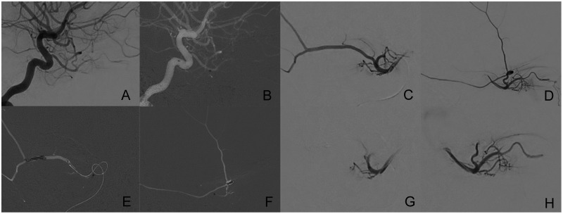
Figure 1.
(a) Lateral digital subtraction angiography (DSA) view of the internal carotid artery and ophtalmic artery. (b) Navigation of the microcatheter into the ophthalmic artery. (c) Anteroposterior and (d) lateral DSA views of the ophthalmic, lacrimal and middle meningeal artery. (e) Anteroposterior and (f) lateral DSA roadmap views after the delivery of the coils in the middle meningeal artery. (g) Anteroposterior and (h) lateral DSA views confirming the occlusions of the middle meningeal artery.

