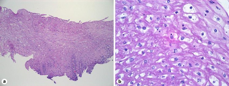
Fig. 2.
a Low power view (100×) of one of the esophagus biopsies, with the basal layer at the bottom and the surface at the top. The epithelium is indistinguishable from normal esophagus, and shows no evidence of hyperkeratosis or dysplasia. There is mild acanthosis (thickening of the epithelium) relative to normal esophagus. b High power view (400×) showing the dark basophilic inclusions and patchy cytoplasmic clearing that can be seen in tylosis patients. These features are also seen in unaffected patients, however, and are of unclear significance.

