Figure 5.
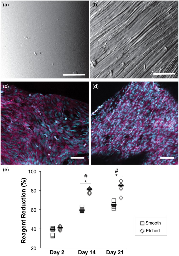
SEM micrographs of scaffold C showing (a) smooth surface texture after printing and (b) unidirectional channels etched using 30 μm particle size sandpaper. Bovine AF cells were cultured in vitro on both types of surfaces for 21 days, stained with Alexa Fluor® 647 phalloidin (magenta) and counterstained with ethidium bromide (nuclei are shown in cyan to enhance contrast). (c) Cells cultured on smooth PCL demonstrated random alignment. (d) Cells cultured on etched PCL had a tendency to align along the underlying surface texture. (e) Alamar blue assay showed significant increases metabolic activity over the culture period, confirming cell proliferation. Scale bars=100 μm. *Statistical significance at single time point; #statistical significance compared with Day 2, P < 0.05

