
Fig. 1.
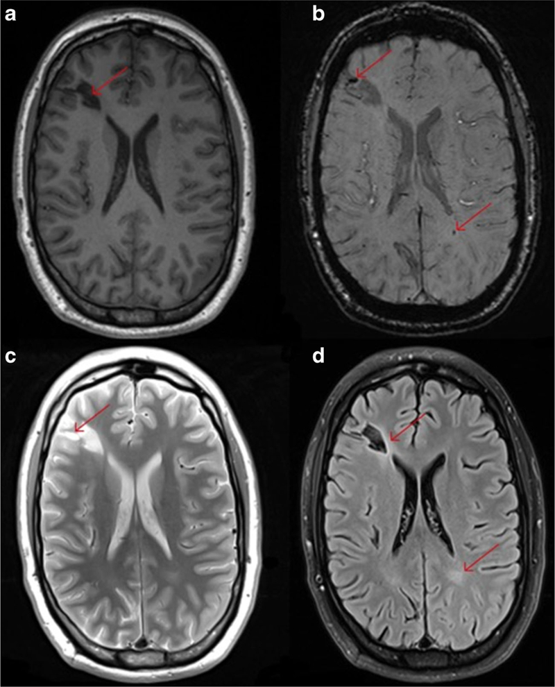
This figure illustrates the different contrast available when using structural MRI to examine a young OIF/OIF Active Duty Service Member diagnosed with a mild TBI (LOC<30 min; AOC<24 h; PTA <24 h). Utilization of multimodal imaging (even structural imaging) can be informative with each sequence potentially adding additional clinically meaningful information about the specific injury incurred. Panel a shows the T1-weighted image and the large hypointense lesion in the right frontal lobe (red arrow). Panel b shows the SWI image and not only shows the large hypointense lesion but several smaller hemosiderin deposits including one in the white matter in the right parietal occipital region (red arrows). Panel c is the T2-weighted image and shows the bright areas indicating inflammation or CSF accumulation around the larger lesion. Panel d is the FLAIR image and shows enhancement around the larger lesion and an area abnormality in the white matter in the left parietal occipital region (contracoup injury)