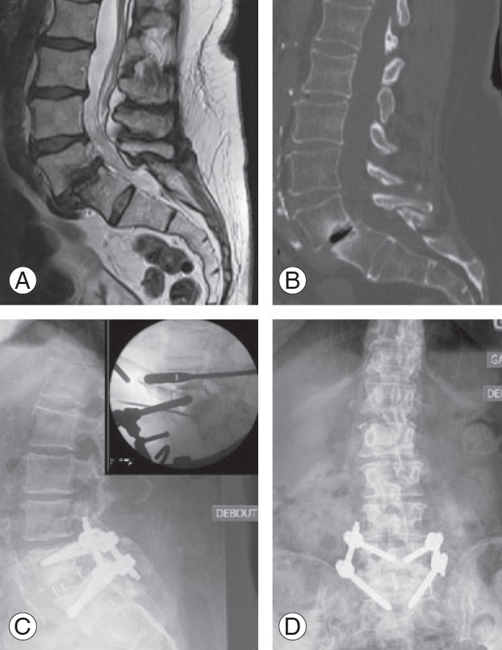
Fig. 2.

Illustrative case of an oblique lumbar interbody fusion procedure. Preoperative sagittal T2-weighted magnetic resonance imaging showing L4–L5 degenerative changes (A) and sagittal computed tomography scan showing grade 1 spondylolisthesis of L4 over L5 (B). Postoperative sagittal (C) and coronal (D) lumbar X-rays showing correction of the spondylolisthesis and improvement of sagittal balance following L4–L5 cage placement and fixation. Insert: preoperative fluoroscopy imaging during placement of the cage in the L4–L5 disk space.
