Figure 3.
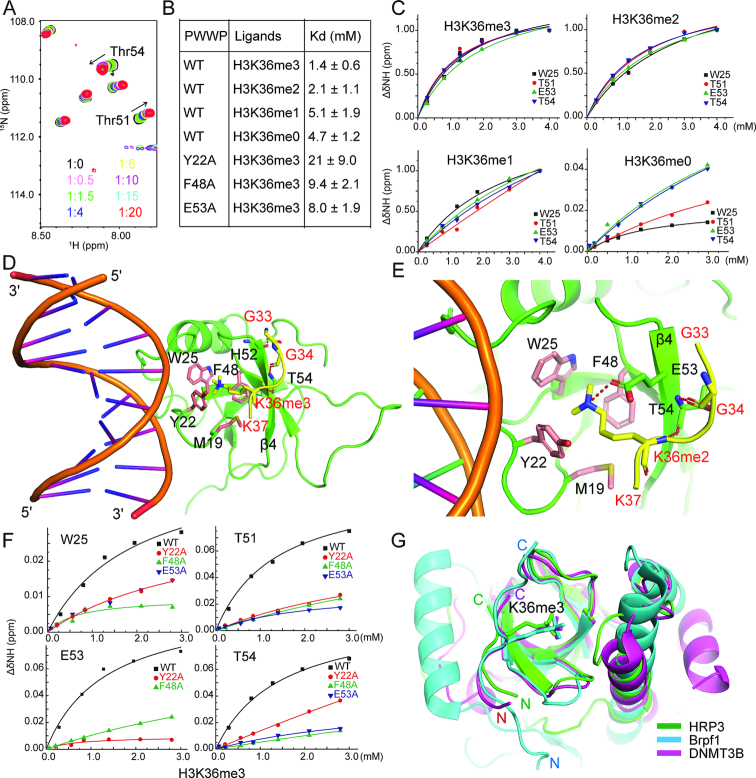
HRP3 PWWP binds to the H3K36me3/2-containing histone peptide. (A) A panel of overlapping HSQC spectra of the HRP3 PWWP domain with various concentrations of the H3K36me3 peptide. (B) A table listing the NMR-based measurements of the dissociation constants for the binding between the wild-type or mutant forms of HRP3 PWWP and various H3K36-containing peptides with/without modifications. (C) NMR-based measurements of different dissociation constants of HRP3 PWWP interacting with H3K36me3/2/1/0-containing histone peptides. (D) Structural details of the ternary complex of HRP3 PWWP/dsDNA/H3K36me3-peptide. The histone peptide is coloured yellow. (E) Structural details of the ternary complex of HRP3 PWWP/dsDNA/H3K36me2-peptide. (F) NMR-based measurements of the dissociation constants for the binding of the different HRP3 PWWP mutants with the H3K36me3 peptides. (G) Overlapped structures of the PWWP domain from HRP3 (in green), Brpf1 (in cyan) and DNMT3B (in magenta) with bound H3K36me3 peptides.

