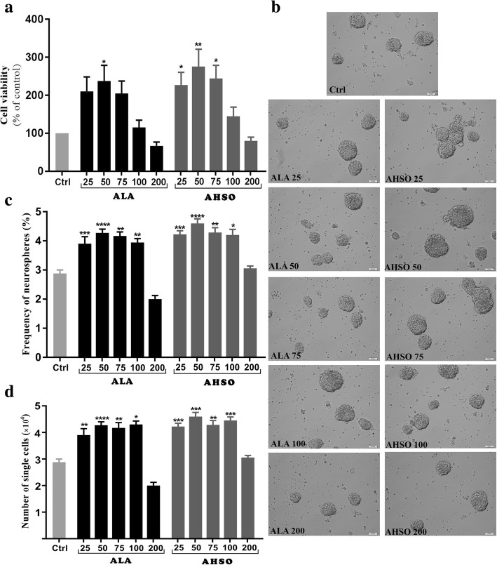
Fig. 1.
Effects of ALA and AHSO on viability and neurosphere formation of eNSCs in vitro. Cells were exposed to different concentrations of synthetic ALA or natural AHSO (25, 50, 75, 100 and 200 μM) for 48 h, then subjected to analysis. a Cell viability assessed by MTT assay. b Representative captures of neurospheres in different groups. Scale bars: 100 휇m. c eNSCs at a density of 500 cells/well were exposed to different concentrations of ALA or AHSO for 5 days. Neurosphere formation was monitored under a microscope and the frequency of neurospheres with a diameter > 50 μm were analyzed. d Cell counts obtained from neurospheres. A one-way analysis of variance (ANOVA) following Tukey post-hoc test was performed to compare the mean values. Data were shown as mean ± SEM. *, p < 0 .05, **, p < 0 .01, ***, p < 0.001 and ****, p < 0.0001 versus control

