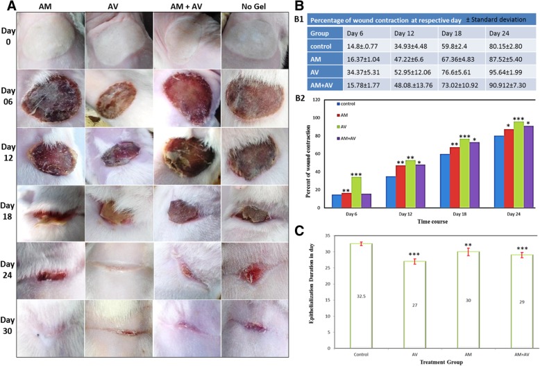
Fig. 5.
Evaluation of wound closure, measurement of wound contraction and re-epithelialization. a Macroscopic wound healing images of Wistar rats at day 0, 6, 12, 18, 24 and 30 which were treated with AM, AV, AM+AV gel or without any gel (control). b Clinical evaluation of wound healing regarding wound contraction in rats at day 6, 12, 18, 24 (b1) and (b2) (level of significant was set * P < 0.05, ** P < 0.01 and *** P < 0.001). c Epithelialization period in different groups (*P < 0.05, ** P < 0.01 and *** P < 0.001)

